

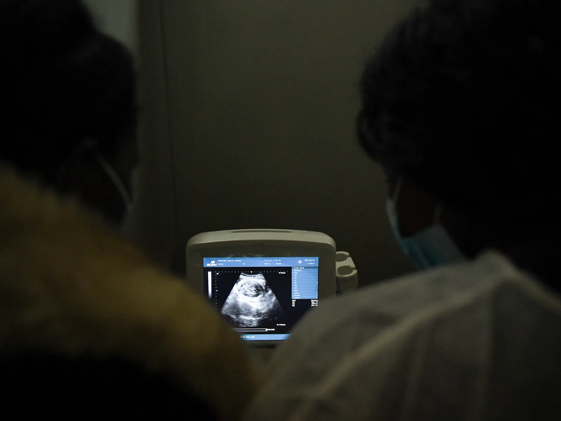
Doctors did not expect that a bleeding in the digestive system of a one-and-a-half-year-old girl would lead to the discovery of an unusual mass inside her stomach.
But subsequent examinations revealed that the mass contained a distorted embryonic structure, in a rare condition known medically as “a fetus within a fetus.”
Bleeding leads to a rare finding
According to a statement by the Ministry of Higher Education and Scientific Research in Egypt, the girl came to the National Liver Institute at Menoufia University, in the north of the country, suffering from gastrointestinal bleeding. She underwent examinations and gastrointestinal endoscopy to determine the source of the bleeding.
During the endoscopy, doctors discovered an unusual mass inside the stomach, and upon completing the medical evaluations, it was found that it contained a deformed fetal structure, in a condition known as “Fetus in fetu,” meaning “a fetus within a fetus.”
These details are consistent with the statements of the institute’s doctors reported by Egyptian media, as they explained that the gastrointestinal endoscopy contributed to discovering the mass, before completing its evaluation and coordination with the surgical team.
Given the child’s young age and the location of the mass, a surgical intervention plan was prepared with the participation of various specialties, and the operation ended with the extraction of the mass and the fetal structure inside it, with the child’s condition stabilizing and her undergoing follow-up after surgery.
How can there be a “fetus” inside a child?
“Fetus within a fetus” is an extremely rare congenital condition that occurs when one twin is trapped inside the body of the other twin during the early stages of fetal development, and then develops abnormally in the form of an embryonic mass of varying degree of morphology.
This formation is not a complete or viable fetus, but may contain, to varying degrees, parts resembling the spine, limbs, or other organs. Organized axial structure, such as the presence of the spine and long bones, are signs that help distinguish the condition from some tumors.
It is likely that the condition is linked to monozygotic twins, but the exact mechanism of its occurrence is still under investigation, and it may sometimes be difficult to distinguish between it and some highly differentiated teratomas, so diagnosis depends on a combination of imaging examinations and histological examination of the mass after its resection.
A rare condition that often appears in the abdomen
“Fetus within a fetus” is an extremely rare congenital condition. A recent systematic review, which included 249 case reports and case series, indicates that most cases appear during the first year of life, and that the retroperitoneum within the abdomen is the most common site. The review also provides a rough estimate of its incidence at about one case per 500,000 live births.
Cases vary in the degree of mass formation and its components, and its diagnosis may not always be clear before surgery. Imaging examinations play an important role in determining its nature and its relationship to surrounding organs, while complete excision and histological examination of the mass represent an essential part of confirming the diagnosis, with favorable results recorded in most published cases.
As for the case of the Egyptian girl, it gains its specificity from the discovery of the mass inside the stomach after it presented itself due to bleeding in the digestive system, before the story ended with its removal and the stabilization of her condition.