

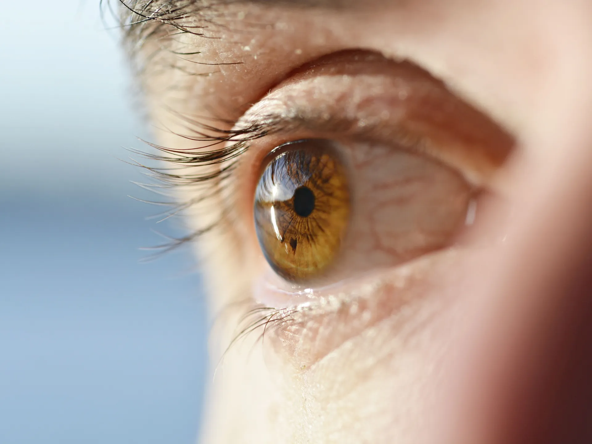
An image of the retina may carry signals beyond vision diseases themselves. A recent study revealed that subtle changes in some layers of the retina are linked to atrial fibrillation, one of the most common heart rhythm disorders, and may appear years before it is clinically diagnosed.
Atrial fibrillation is a disorder in the electrical activity of the upper chambers of the heart, i.e. the atria, leading to an irregular pulse and ineffective contraction.
This may allow blood to stagnate, especially within the left atrial appendage, which increases the possibility of the formation of clots that can travel to the brain and cause a stroke.
The US Centers for Disease Control and Prevention indicates that atrial fibrillation is associated with a nearly 5-fold increase in the risk of ischemic stroke after taking into account usual risk factors.
Estimates of the global burden of disease indicate that atrial fibrillation and atrial flutter were affecting about 52.55 million people around the world in 2021. One of the problems with atrial fibrillation is that it may pass without symptoms or with unclear symptoms, despite its association with an increased risk of stroke, heart failure, and other complications.
A recent study – published in the journal “PLOS Digital Health” on September 10 – showed that 62% of people with atrial fibrillation were not aware that they had it before they were diagnosed, and revealed that changes that occur in the layer of cells located in the back of the eye may help detect atrial fibrillation up to 4 full years before it is diagnosed.
What did the study see in Al Ain?
The researchers analyzed retinal imaging data from more than 90,000 people in two large British cohorts. The first – the AlzEye base affiliated with Moorfields Eye Hospital – included 50,651 people, including 5,375 people who had atrial fibrillation.
As for the second group, it came from the British Biobank (UK Biobank), and included in the future analysis 39,13 people who did not have atrial fibrillation at the beginning of follow-up, and then 838 of them developed the disease during an average follow-up of 4 years.
The researchers relied on optical coherence tomography (OCT), which is a non-invasive examination that uses light to produce high-resolution images of the retinal layers, along with color images of the fundus of the eye.
They found that people with atrial fibrillation had changes in a number of retinal layers, and the most consistent finding was a thinning of the macular ganglion and inner plexiform layer (mGCIPL), a neural layer important in transmitting visual information from the retina toward the brain.
What is most striking is that people who did not have atrial fibrillation when examined, and were subsequently diagnosed with it, had insomnia on average about 4 years before the clinical diagnosis.
After taking into account factors such as age, gender, blood pressure, diabetes, smoking and alcohol, each standard decrease in the thickness of this layer was associated with a 27% increase in the subsequent incidence of atrial fibrillation.
However, the study does not mean that retinal thinning leads to fibrillation, nor that an eye examination can diagnose it alone. Rather, it reveals a connection that may help in the future to identify people who need additional cardiac examinations.
How does the eye reveal what is happening in the heart?
Dr. Ihab Saad Othman, Professor of Ophthalmology and Surgery at Cairo University Faculty of Medicine, told Al Jazeera Net that the retina is directly linked to the central nervous system, and its tiny blood vessels give doctors a unique window to see the effect of systemic diseases on the blood circulation.
He points out that OCT devices can measure the thickness of the retinal layers with a high degree of accuracy, which has made them an essential tool in diagnosing and monitoring eye diseases, such as glaucoma, retinal diseases, and the optic nerve.
The retina consists of multiple interconnected layers that transmit the signal from light-sensitive cells to neurons that collect visual information and send it through the optic nerve to the brain.
Therefore, the new result is important, as the presence of a regular change in the thickness of some nerve layers in people with atrial fibrillation raises the possibility that the eye may be able to show an early effect of disturbances occurring in the blood circulation before the disease appears clinically.
Dr. Khaled Farouk, Professor of Critical Care Medicine at Kasr Al-Aini Faculty of Medicine, Cairo University, points out that atrial fibrillation may be intermittent, appearing and then disappearing, and therefore the ECG may be normal if it is performed at a time when a seizure does not occur.
He added to Al Jazeera Net that some infected people do not feel symptoms, while others may suffer from palpitations, fatigue, shortness of breath, dizziness, or chest pain, which are symptoms that are not limited to this disease alone.
A disease that may remain silent
The difficulty of detecting atrial fibrillation does not mean that all sufferers are unaware of their condition, but lack of awareness of the disease is a clear problem.
In a separate survey conducted by the American Heart Association in 2025, which included 770 patients with atrial fibrillation, 62% of them said that they did not know about the disease before they were diagnosed with it. This percentage does not come from a study of the retina itself, but rather from the association’s independent awareness-raising research.
The importance of early detection stems from the fact that treatment may include medications to regulate heart rate or rhythm, in addition to anticoagulants for patients who need them to reduce the risk of clots and stroke.
Do small clots leave traces in the retina?
Researchers suggest a possible explanation for the thinning of the nerve layers in the retina, which is that circulatory disturbances or micro-blockages may reduce blood supply to the retinal cells, which over time leads to the loss of some nerve tissue.
But this explanation is still a hypothesis that requires further research, and the study did not prove that microclots are the direct cause of the changes it observed.
There are other indications that support a relationship between the eye and atrial fibrillation. A previous study of 169 people with AF found that the presence of retinal ischemic perivascular lesions (RIPLs) was associated with a history of stroke, even after adjusting for some known risk factors, with an odds ratio of 2.59.
But this result also does not mean that these lesions alone predict who will suffer a stroke, as the study was cross-sectional and relatively small. Rather, it adds evidence that the small vessels in the retina may carry information about the state of blood circulation in the rest of the body.

Eye examination is not a substitute for an ECG
Dr. Khaled Farouk believes that the link between the retina and atrial fibrillation makes sense from a biological standpoint, because the retina is part of the central nervous system, and its tiny vessels may reflect changes occurring in the body’s vessels and blood circulation.
But he stresses that retinal examination cannot currently replace electrocardiography (ECG) or heart rhythm monitoring methods used to confirm the diagnosis.
This is consistent with the researchers themselves, who explained that the potential goal of eye examination is not to diagnose atrial fibrillation directly, but rather to use it as an aid to identify people who may need more accurate heart examinations.
Will the eye clinic become a station for detecting heart diseases?
Dr. Ihab Saad Othman says that the results open an important door, especially in people who combine risk factors such as high blood pressure, diabetes, smoking, and atherosclerosis.
But he stresses that turning a research observation into a general recommendation requires additional studies that determine who should be screened and what value OCT adds over traditional risk factors.
Dr. Khaled Farouk points to other challenges, including standardizing the method of measuring retinal layers between different devices, proving that adding the examination to traditional cardiac evaluation improves the ability to detect the disease or prevent its complications, and studying the economic feasibility of including it in screening programs.
This point is important; There is currently no recommendation that all people have an OCT scan for atrial fibrillation.
When should you see a doctor?
If symptoms such as frequent palpitations or fluttering in the chest, irregular pulse, unexplained shortness of breath, dizziness, unusual extreme fatigue, or chest pain occur, a medical evaluation should be sought.
Chest pain, fainting, or signs of a stroke – such as sudden weakness in the face or arm or difficulty speaking – require urgent medical care.
The most important message presented by the new study is that the eye may in the future carry information about the heart that we were not previously searching for.
An examination that takes only minutes in an eye clinic may not diagnose atrial fibrillation, but it may one day help draw attention to a patient whose heart needs to be monitored before the problem becomes more serious.