


In the history of medicine, major leaps often began when scientists were able to see what was hidden inside the body. In 1991, a research team from the Massachusetts Institute of Technology (MIT) and collaborators published a new technique that made it possible to produce high-resolution cross-sectional images of biological tissue using light, called optical coherence tomography (OCT).
The first study showed images of tissues, including the retina of a human eye from a cadaver and a coronary artery, before researchers succeeded in 1993 in using the technology to photograph the retinas of living people, opening the door to one of the most important transformations in the history of diagnosing eye diseases.
After more than 3 decades, the names of 3 of the most prominent developers of the technology, James Fujimoto, David Huang, and Eric Swanson, returned to the forefront with the Claveret Foundation including them on the Citation Laureates list for the year 2026, in recognition of the development of optical coherence tomography and its medical and research uses.
Claveret’s list does not mean that scientists are officially nominated for the Nobel Prize, as the actual nominations remain secret, and the foundation itself confirms that it does not predict a winner for a particular year, but it selects scientists that it believes the impact of their research puts them at the level of achievements that the Nobel has previously rewarded.
As the 2026 Nobel Prize in Medicine and Physiology is announced, the technology raises an exciting question: Will OCT catch up with other medical imaging technologies whose developers previously led to the Nobel Prize, such as computed tomography and magnetic resonance imaging?
How does light see inside the eye?
The idea of optical coherence tomography can be simplified by analogy to ultrasound, but instead of sending sound waves and receiving echoes, the technique relies on light.
But the main obstacle is that sound is relatively slow, and its return time from tissues can be measured, while light moves at a speed of approximately 300,000 kilometers per second, which makes measuring its return time directly from small distances within tissues extremely difficult.
To overcome this, the technology relies on the principle of “optical interference”, where the light inside the device is divided into two paths: one of them heads towards the eye or tissue to be imaged, while the other follows a known reference path.
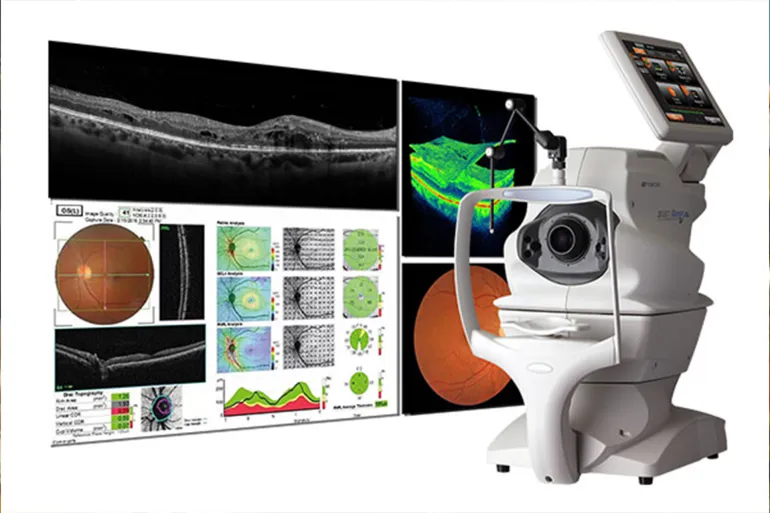
When the scattered light from the tissue returns and meets the light coming from the reference path, an interference pattern is generated that a computer can analyze to extract accurate information about the positions of different structures in depth.
By collecting thousands of measurements, the device can build a high-resolution cross-sectional image of the retina in a few seconds. Its accuracy in some modern systems reaches a few microns, as the doctor can see a large number of layers of the retina and its delicate structures inside the living eye, without surgery or direct contact with the tissue.
From the surface of the retina to its depths
Before the advent of OCT, a fundus examination allowed the doctor to see the surface of the retina and optic nerve, but it did not provide the detailed cross-sectional image that revealed what was happening within the layers of tissue itself.
With the introduction of technology into clinics, it became possible to measure retinal thickness, monitor fluid accumulation, and follow microscopic changes objectively and repeatedly.
The importance of this technology is no longer limited to detecting the disease, but rather extends to monitoring its development, measuring the patient’s response to treatment, and making more accurate decisions regarding the timing of medical intervention.
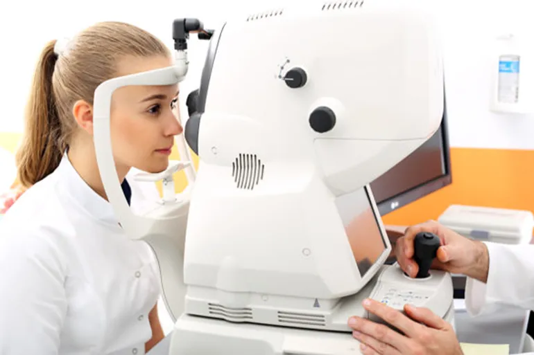
What has changed OCT in ophthalmology?
- Age-related macular degeneration: The disease is one of the main causes of vision loss in the elderly, and its early stages may pass without obvious symptoms. OCT allows monitoring minute changes in the retina, fluids, deposits and structures associated with the disease, and also plays an essential role in monitoring response to treatment.
- Diabetic retinopathy: The disease may lead to fluid leakage into the retina and the occurrence of diabetic macular edema in the center of vision. OCT allows you to measure retinal thickness and monitor fluid accumulation with micron accuracy, then monitor changes after treatment, including intraocular injections.
- Glaucoma: The technique can measure the thickness of the layer of retinal nerve fibers and ganglion cells associated with the optic nerve, and in some patients it may detect early structural changes before visual field loss becomes apparent on functional tests.
Farther than the eye
OCT does not stop at ophthalmology, but its applications have extended to other specialties. In cardiology, intravascular versions of the technology are used to obtain high-resolution images of the walls of coronary arteries, help evaluate plaques, determine the dimensions of lesions, guide the implantation of stents, and ensure that they expand and adhere properly to the artery wall.
The technology is also being used and developed in dermatology, gastroenterology, oncology and surgery, with the aim of obtaining microscopic images of tissues without cutting them.
But these uses do not mean that OCT has become a general alternative to biopsy or traditional diagnostic methods, as many of its applications outside ophthalmology are still complementary to other tests or are under development.
3 minds behind the technology
OCT is the result of a clear intersection between physics, engineering, and medicine. James Fujimoto led the laboratory at MIT from which a key part of the work was initiated, and he and his team helped develop the use of low-coherence light to produce high-resolution images of tissue.
As for David Huang, who was then a student in a joint medical and research program between Harvard and the Massachusetts Institute of Technology (MIT), he was the first researcher in the detailed study published in the journal “Science” in 1991, which introduced the technology to the world.
Eric Swanson comes from optical communications at MIT Lincoln Laboratory, and his expertise in fiber optics and optoelectronics has contributed to the development of faster and more usable systems.
The transfer of technology from the laboratory model to commercial devices helped open the way for its wide spread in eye clinics around the world.
Tens of millions of tests
Within a few years, OCT has gone from a laboratory experiment to an essential tool in modern ophthalmology. Published estimates indicate that about 30 to 40 million OCT examinations are performed annually around the world, which reflects their widespread use in diagnosing and following diseases of the retina and optic nerve.
The technology has helped detect diseases at more treatable stages, monitor their development quantitatively, and follow up on patients’ response to modern treatments, especially in macular degeneration, diabetic retinopathy, and glaucoma.
Its importance lies in the fact that it not only provided a clearer picture, but also changed the method of monitoring the disease itself, as the doctor could now compare tissue thickness, fluid accumulation, and structural changes over time with an accuracy that is difficult to achieve with traditional examination alone.
From Lasker to Nobel?
In 2023, Fujimoto, Huang, and Swanson received the Lasker-DeBakey Clinical Research Award for the invention of OCT, one of the most prestigious medical awards in the world.
The three were also awarded the US National Medal of Technology and Innovation for the year 2022, and received it from former US President Joe Biden at the White House in October 2023.
In September 2026, the Claveret Foundation included them in this year’s Citation Laureates list, in recognition of the development of optical coherence tomography as a new imaging modality in medicine and biomedical research.
Since the launch of the Claveret program in 2002, a number of scientists previously selected by the Foundation have subsequently won the Nobel Prize, but inclusion on the list does not mean an official nomination and does not represent a certain expectation of winning.
However, the connection to the Nobel seems logical from the perspective of the history of medicine, as the prize has previously been awarded for achievements that changed the way the body is viewed from the inside, including computed tomography in 1979 and magnetic resonance imaging in 2003.
Light is a diagnostic method
OCT was not just a technical addition to ophthalmic equipment, it provided doctors with a way to view living tissue at near-microscopic resolution, with the ability to repeat the examination and compare results over months and years.
Thus, the retina was transformed from a surface that the doctor could look at into a tissue whose layers could be measured and whose changes could be monitored moment by moment.
Whether Fujimoto, Huang and Swanson’s journey to the 2026 Nobel Prize ends or not, the impact of the technology is already well established in medical practice. Every time OCT detects tiny fluid inside the retina or early damage to the optic nerve, the impact of an idea that began more than 3 decades ago with a single attempt is embodied: using light to see what was not visible.