
The results of a new study pave the way for expanding the scope of the use of a trial device to open the brain blood barrier, with the aim of improving treatments and diagnosis for patients with brain tumors and other diseases of the nervous system.
The study identified a mark of sound emissions as a reliable scale to determine the opening of the brain blood barrier, and presented the first technical description of this use.
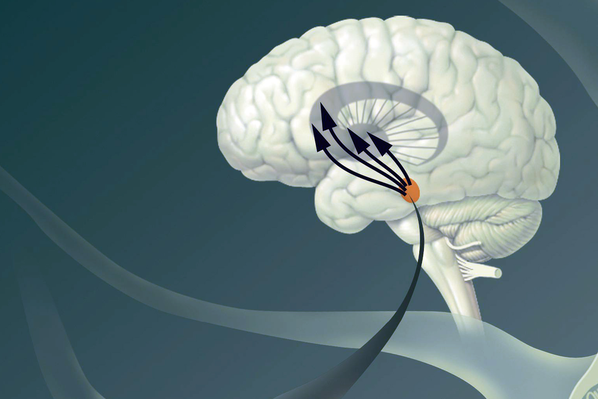
The blood hemorrhagic barrier is a specialized network of vascular and brain cells that act as a brain safety system to protect against the invasion of toxins and dangerous microbes, but also reduces the effectiveness of treatments in the brain, such as chemotherapy medications.
The study was conducted by researchers from the University of Maryland Medical College, Brigham Hospital and Women in Boston and other institutions in North America, and the results of the study were published in Device Journal on August 25, and the Yorik Alrt website was written about.
Microscopic bubbles filled with inert gas in the patient’s bloodstream are injected, and are activated by ultrasound energy in the targeted area of the opening of the brain septum, while monitoring all of this by magnetic resonance imaging to determine effectiveness.
“When these microscopic bubbles are raised with low -severity ultrasound, fluctuating within the energy field, causing temporary mechanical disorders in the walls of the blood vessels,” said Dr. Pavloos Anastasiadis, Assistant Professor of Neurosurgery at Michigan State University in the United States and the author of this study.
The researchers found that the frequency sound waves captured as signs of sound emissions resulting from circular accurate bubbles are a reliable measure of predicting the opening of the brain blood barrier in patients.
Safe and practical technique
It is expected that the methods and analyzes described in the study will allow researchers and doctors to monitor the treatments of the blood barrier opening, their comparison and description through various devices, patients and environments in the future, which helps to facilitate and determine the location of the delivery of the drug in the brain.
“In light of the increasing availability of multiple devices to connect ultrasound energy, varies the standards of treatment, and the difference in immediate monitoring and control methods, there is an urgent need that has not yet been able to determine and unify the treatments of the blood barrier opening with concentrated ultrasound,” said Dr. Woodworth.
“Monitoring sound emissions and doses systems derived from them allows an opportunity to unify the concept of concentrated ultrasound, and the data and analyzes presented in this study contribute to developing this systematic model and the field of concentrated ultrasound.”
Doctors, scientists, and engineers led by Dr. Graim and Woodworth, Professor and Head of the Department of Neurosurgery at the University of Maryland, and director of the program of treatment and research of brain tumors at the Marilyn Center and Stoyart Greenpom, comprehensive cancer collection, collected data from 34 patients with a dibbian tumor, each receives up to 6 monthly treatment courses that include a total of 972 individual sessions of concentrated ultrasound To determine the most effective use of concentrated ultrasound to achieve a reliable opening of the cerebral hemorrhoids.
Dr. Alexandra J. Gulbe, Director of Neurosurgery, directed with pictures at Brigham Hospital, Women, and the main author of the study.
“Our work depends on these discoveries, as we find a safe and practical technique that allows us to open the brain blood barrier frequently in patients with diet aromatic tumor before chemotherapy,” she added.